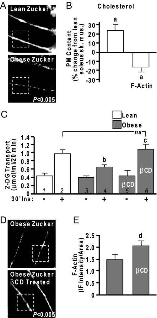
Figure 9.
Cholesterol Lowering Normalizes Insulin Sensitivity in Obese Zucker Skeletal Muscle
Rat epitrochlearis muscle (A) and soleus muscle (B–E) from lean/obese Zucker rats were labeled with antibodies against F-actin, imaged by confocal microscopy (A and D), and digitally quantitated using MetaMorph software (B and E). Remaining soleus muscle was fractionated for PM cholesterol analyses (B). Contralateral soleus muscles were subjected to basal and insulin-stimulated 2-DG uptake measurements (C) as described in Materials and Methods. A subgroup of these muscles was exposed to 2.5 mm βCD for 30 min before the 30-min insulin stimulation (30′Ins). Values are means ± sem from five independent experiments. a, P < 0.05 vs. lean; b, P < 0.05 vs. lean +Ins; c, P < 0.05 vs. obese +Ins; d, P < 0.05 vs. obese −βCD. ns, Not significant; sk. mus., skeletal muscle.

