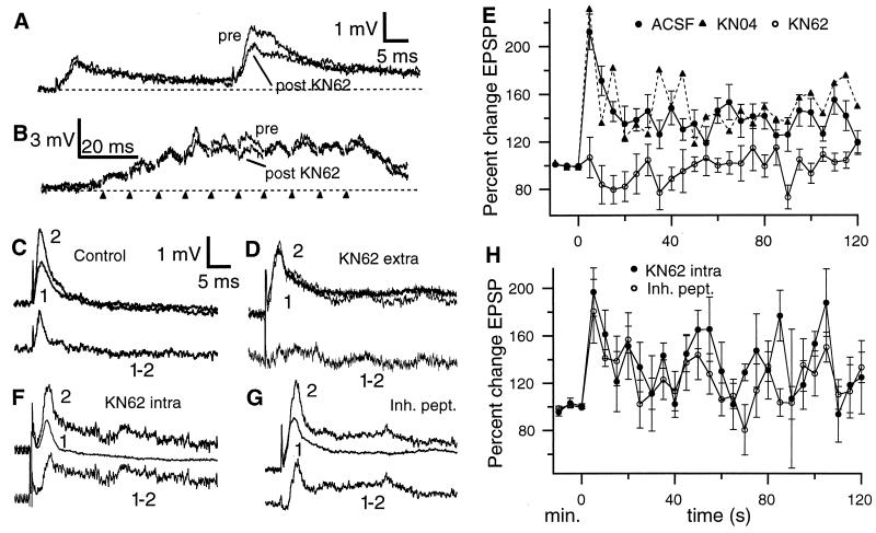
Figure 2.
(A) Paired pulse facilitation of StF (50 ms interval). StF stimulation evokes a rapid EPSP with a slow decay phase; the second pulse evokes a substantially larger EPSP. KN62 does not alter the first EPSP (superimposed trace from the same cell 15 min after drug ejection) but reduces PPF. The stimulus artefact was digitally removed in this and subsequent figures. (B) Effect of KN62 on train stimulation of StF. Although a reduction of some EPSPs does occur under KN62, this is slight due to the strong voltage dependent augmentation of later EPSPs in the train, so that the overall response to tetanic stimulation is not grossly altered. Markers indicate times of stimulation. (C, Upper) Superimposed pre- and posttetanus (5 s) EPSPs under control (ACSF) conditions. (Bottom) Subtraction of pre- from posttetanus EPSP reveals extent of PTP. (D, Upper) superimposed pre- and posttetanus (5 s) EPSPs after application of KN62. (Bottom) Subtraction of pre- from posttetanus EPSP reveals block of PTP. (E) Time course of PTP with ACSF and KN04 (error bars for KN04 plot are omitted for clarity); KN04 cases are indistinguishable from control. KN62 completely blocks PTP at all posttetanus times. (F and G, Upper) Superimposed pre- and posttetanus (5 s) EPSPs after intracellular application of KN62 (F) or a CaMK2α inhibitory peptide (G). (Bottom) subtraction of pre- from posttetanus EPSP reveals normal PTP. (H) The time course of PTP is not altered by intracellular application of CaMK2α antagonists. Note that in E and H the pretetanus time scale is in minutes (pretetanus EPSPs were evoked at 5-min intervals to assess their stability) while the posttetanus scale is in s.

