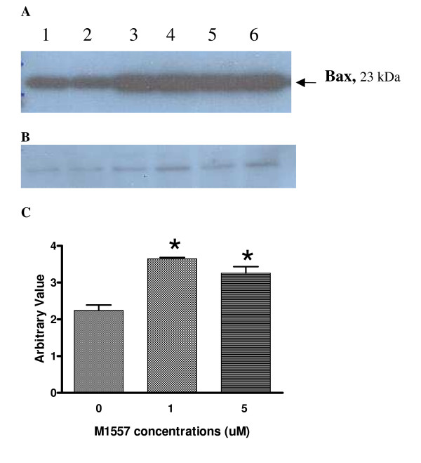
Figure 5.
Western blotting was used to detect pro-apoptosis Bcl2-associated X protein (Bax) in M1557 peptide treated and untreated cells (A). After treatment with M1557 peptide at 2 μM and 5 μM concentrations, the level of Bax increased significantly compared to that in the untreated cells (P < 0.01) (C). (B) The level of β-actin in each sample was detected by a polyclonal anti-actin antibody (Sigma, UK). β-actin was used as internal standard to ensure the equal amounts of protein was loaded.

