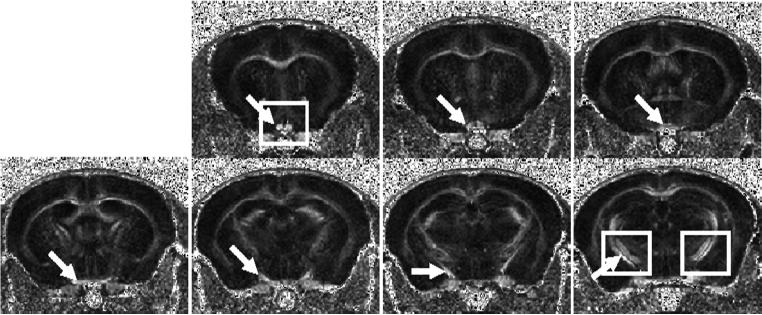
Figure 2.
Contiguous slices of RA maps covering the optic nerve, chiasm, and optic tract acquired from a normal mouse. The arrow identifies the optic nerve (the first 2 slices), optic chiasm (the 3rd and 4th slices), and optic tracts (the 5th through 7th slices). The expended views of optic nerve and tract specified by the rectangles are expanded in Fig. 3.

