
Figure 1.
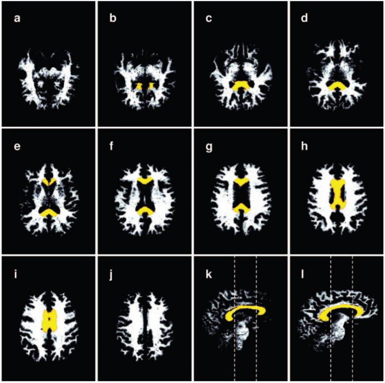
A series of segmented white matter images in the native space of the T1-weighted images from which they were calculated. The corpus callosum (CC) region of interest is superimposed in color. Volume measurements of the CC were performed by applying the CC masks to these white matter images. The CC ROI has been morphed from the MNI space where it was defined to the native space of the T1 image prior to application on the fractional anisotropy images. (a-j) Axial slices in an inferior to superior direction. (k) Paramedian sagittal slice offset 2 mm from the sagittal plane. (l) Sagittal slice offset 10 mm from the sagittal plane. Dashed lines in k-l indicate the caudal and rostral limits of the body of the CC.