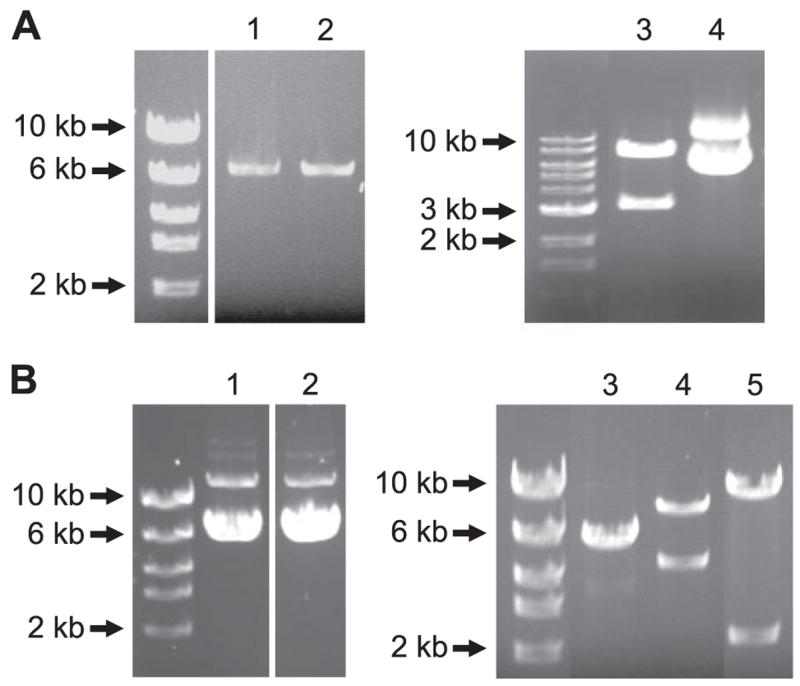
FIGURE 3. Full-length CaV1.2 cDNAs cloned from rat small cerebral arteries.

A, full-length CaV1.2e1b and CaV1.2e1c were cloned by RT-PCR and sub-cloned into pGEM-T Easy vector. Lane 1, CaV1.2e1b; lane 2, CaV1.2e1c; lane 3, CaV1.2e1c was released from pGEM-T easy vector by NotI digestion; lane 4, pGEM-T easy-CaV1.2e1c. B, CaV1.2e1b and CaV1.2e1c were subcloned into pIRES-hrGFP II vector, and the orientation direction of CaV1.2 in the expression vector was revealed by EcoRV digestion. Lane 1, pIRES-CaV1.2e1c-hrGFP II; lanes 2 and 3, pIRES-CaV1.2e1b-hrGFP II; lane 4, EcoRV digestion product of pIRES-CaV1.2e1b(+)-hrGFP II; and lane 5, EcoRV digestion product of pIRES-CaV1.2e1b(−)-hrGFP II.
