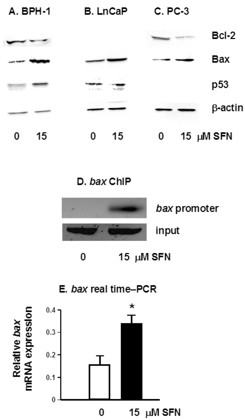
Fig. 4.

SFN alters the expression of pro- and anti-apoptotic proteins. Attached cells were harvested 48 h after treatment of BPH-1 (A), LnCaP (B) and PC-3 cells (C) with 0 or 15 μM SFN, and cell lysates were immunoblotted for Bcl-2, Bax and p53, as indicated. Equal protein loading was confirmed using β-actin. Results are representative of two or more separate experiments. (D) BPH-1 cells were treated with 0 or 15 μM SFN and ChIP was performed as described in the legend to Figure 3, except that PCR was performed with primers to the bax promoter. (E) Real-time PCR results for bax mRNA expression in BPH-1 cells 12 h after treatment with 0 or 15 μM SFN; mean ± SD, n = 3; *P <0.05.
