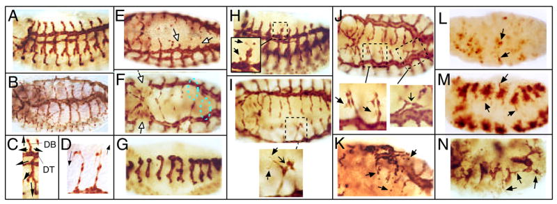
Figure 4.
Tracheal defects in Dakt1 and pnt mutants. Embryos were stained with mouse monoclonal anti-β-Gal antibody. In all panels, anterior is to the left. (A) Lateral view of a stage 14 1-eve-1 embryo, representing wild type. (B) Dorsal-lateral view of a stage 16 1-eve-1 embryo, showing the fully formed tracheal system. (C) One wild-type tracheal metameric subunit at stage 14. Migratory paths of the six primary branches are marked by arrows. Two relevant branches are indicated: DB, dorsal branch; DT: dorsal trunk. (D) Close-up view of two wild-type dorsal branches at stage 16. Arrows indicate the directions of the extending cellular processes at the tips of the elongating tubes. (E) Dorsal-lateral view of a stage 15 pntΔ88, 1-eve-1/+ embryo. Empty arrows point to "stunted" dorsal branches in which tracheal cells fail to migrate out. (F) Dorsal view of a stage 16 pntΔ88, 1-eve-1/+ embryo. Open arrows point to the gaps in the dorsal trunks and dashed circles marked positions where dorsal branches are missing. (G) Lateral view of a homozygous stage 15 pntΔ88, 1-eve-1 embryo. Very little branching occurs. (H) Lateral view of a stage 13 Dakt1q, 1-eve-1/+ embryo. Arrows in the enlarged view point to ectopic filopodia and an errand cell sprouting from the main dorsal trunk. (I) Dorsal view of a stage 16 Dakt1q, 1-eve-1/+ embryo. Arrows in the enlarged view point to ectopic filopodia. Sharp arrow points to the ectopic connection between two adjacent subunits. (J) Dorsal view of a homozygous stage 16 Dakt1q, 1-eve-1 embryo. Arrows in the enlarged view point to ectopic filopodia. Sharp arrow points to the ectopic connection between two adjacent subunits. (K) Dorsal view of a homozygous stage 16 Dakt1q, 1-eve-1 embryo. Arrows point to ectopic branches and breakaway tracheal cells. (L) Lateral view of a stage 14 Dakt1q, 1-eve-1/pntΔ88, 1-eve-1 embryo. Arrows point to ectopic branches and breakaway tracheal cells. (M) Ventral view of a stage 13 Dakt1q, 1-eve-1/pntΔ88, 1-eve-1 embryo. Most of the tracheal cells remain within the tracheal placodes while erratic branches are seen sprouting out (arrows). (N) Lateral view of a stage 13 Dakt1q, 1-eve-1/pntΔ88, 1-eve-1 embryo. The tracheal subunits lack stereotypical branching, similar to the phenotype observed in the pnt mutant (G) but erratic small branches are seen sprouting out (arrows).

