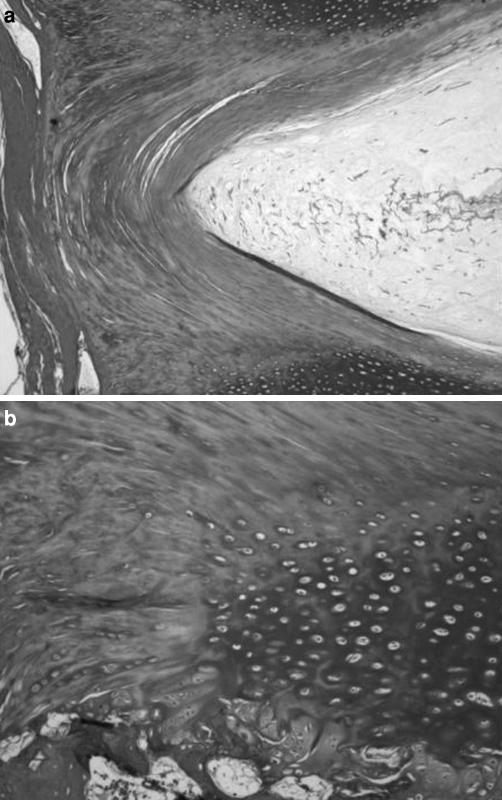
Fig. 7.
Annular fibers adjacent to the longitudinal ligaments anchored to the mineralized cartilaginous endplate zone. A mineralized tidemark line delimited the lateral portions of the mineralized endplate zone. Mineral deposition stripes from the mineralized cartilaginous endplate zone followed the initial portion of fibers of the annular fibers adjacent to longitudinal ligaments and of the fibers continuing in the longitudinal ligaments. Numerous oval chondrocytes were present between the mineral deposition stripes; a Masson’s trichrome, ×4; b Masson’s trichrome, ×10

