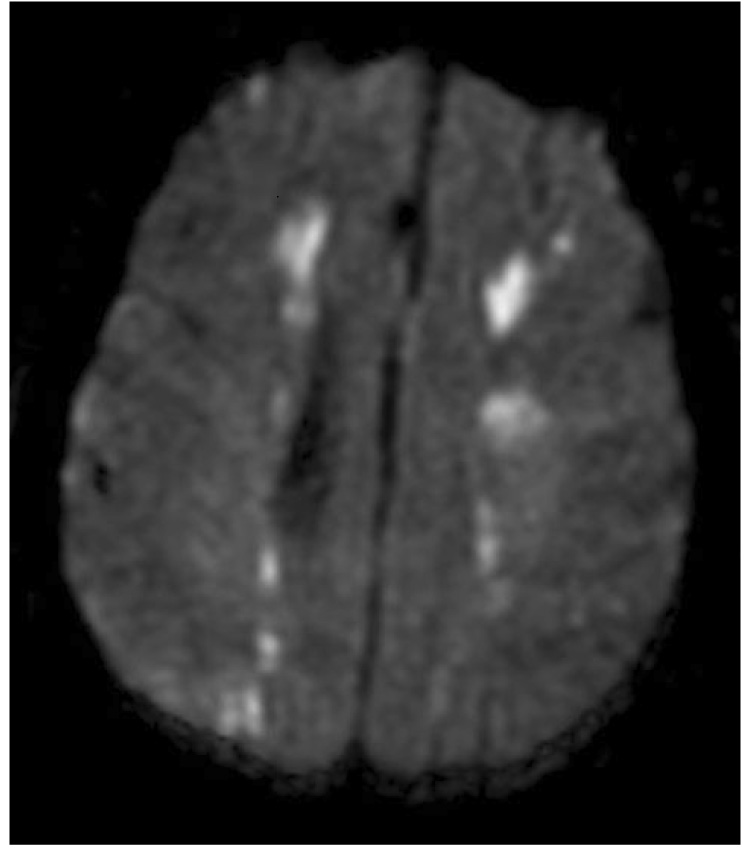
Figure 1.

Diffusion weighted brain magnetic resonance image of a patient with stroke after cardiac surgery. The bright images represent brain ischemic injury. The location of the injury in the end vascular territory of the anterior and middle cerebral arteries and the middle and posterior cerebral arteries is consistent with a watershed infarction due to cerebral hypoperfusion.
