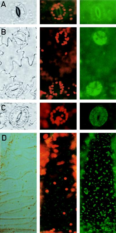
Figure 3.
Detection of GFP in tobacco leaf epidermic tissue. Transmission (Left column) and epifluorescence microscopy to visualize either the red chlorophyll fluorescence and the green GFP fluorescence (Center column) or only the GFP fluorescence (Right column). Stomatal guard cells from control, untransformed tobacco plant (A), plant transformed with pBI-GFP5 (B), lacking the targeting sequence, and plant transformed with pBI-TP-GFP5 (C), containing the targeting peptide. (D) A hair cell of a plant as described in C. (10 μm = 3.2 mm).

