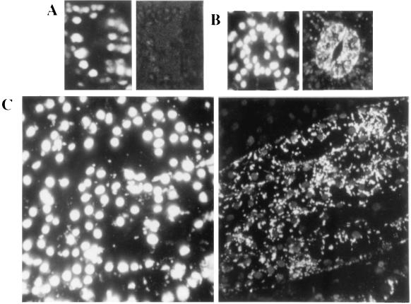
Figure 6.
Confocal images of tobacco leaf epidermic tissues. Projection of 43- to 58-serial sections taken at 1-μm intervals; Left column, images obtained in the red channel (chlorophyll fluorescence); Right column, images visualized in the green channel (GFP fluorescence). Stomatal guard cells of a control, untransformed tobacco plant (A), stomate (B), and epidermic tissue (C) of a tobacco plant transformed with pBI-PT-GFP5, containing the targeting peptide. Mitochondria are observed in the green channel. (10 μm = 4 mm).

