
Figure 1. Loss of PUMA activates cell cycle checkpoints through p21.
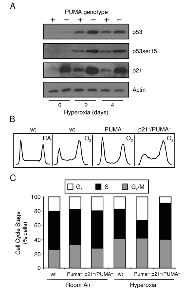
(A) Expression of p53, p53 ser-15 and p21 in HCT116 wt and PUMA− cells exposed to a hyperoxic time course with actin used as a loading control. Immunoblots were representative of three separate experiments. (B) Representative DNA histograms of HCT116 wt, PUMA− and p21−/PUMA− cells exposed to 4 days of room air (RA) or hyperoxia (O2). (C) Mean average of HCT116 wt, PUMA− and p21−/PUMA− cells in G1, S and G2/M exposed to 4 days of room air or hyperoxia (n=3).