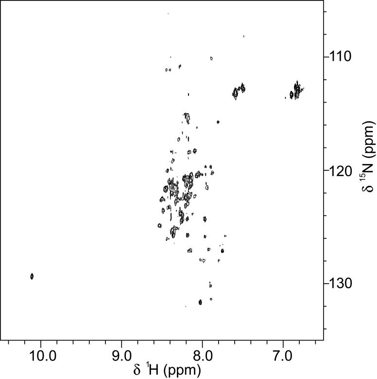
Fig. 3.
NMR analysis of AID. Two-dimensional 1H-15N HSQC spectrum of uniform 15N-isotopic labeled AID. The final concentration of AID was 50 μM in 10 mM phosphate buffer, pH 7.3, containing 100 mM NaCl and 5% D2O. Spectra were collected at 25 °C with a Bruker DMX500 NMR spectrometer (500 MHz for protons) equipped with a triple resonance cryoprobe.

