
Figure 1.
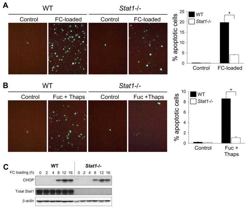
SRA/ER stress-induced macrophage apoptosis requires STAT1. ( A–B)Peritoneal macrophages from wild-type (WT) or Stat1−/− mice were incubated for 17 h with medium alone (Control) or medium containing acetyl-LDL plus the ACAT inhibitor 58035 (FC-loaded); or 21 h with medium alone (Control) of medium containing 50 μg/ml fucoidan and 0.5 μM thapsigargin (Fuc + Thaps). Mid- and late-stage apoptosis were assessed by staining with Alexa Fluor 488-conjugated annexin V (green) and PI (orange), respectively. Representative merged fluorescence and bright-field images and quantitative data from 3 fields of cells for each condition are shown. *, p = 0.001 by Bonferroni after ANOVA. (C) Lysates from wild-type and Stat1−/− macrophages were FC-loaded for the indicated times and subjected to immunoblot analysis to detect CHOP, total STAT1, and β-actin.