
Figure 6.
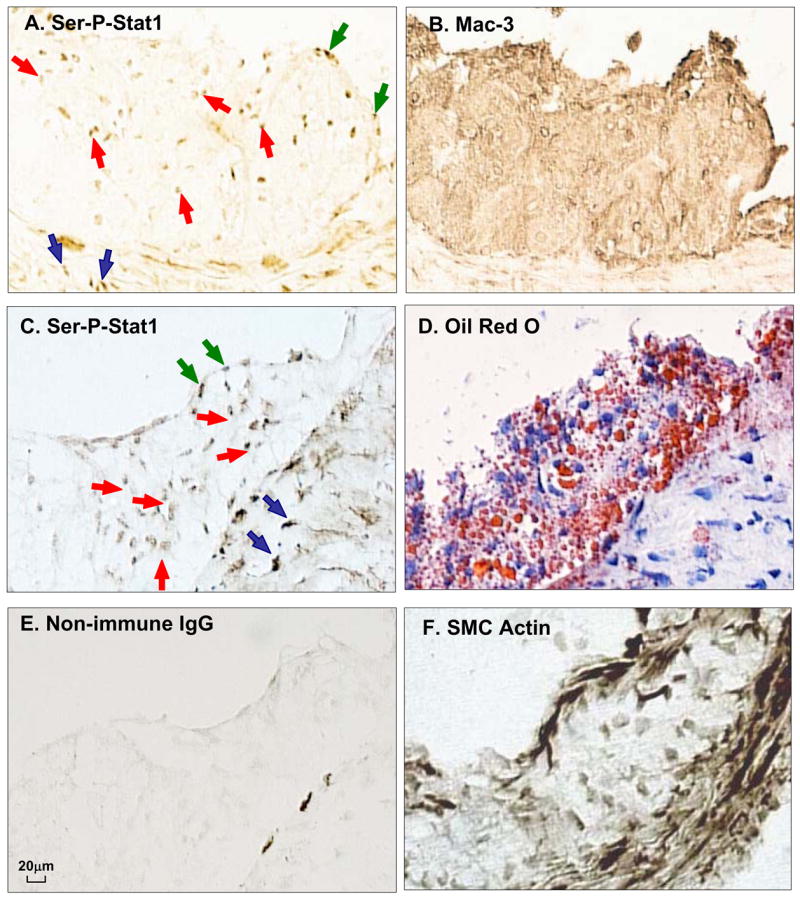
STAT1 is serine-phosphorylated in atherosclerotic lesions from Ldlr−/− mice. Adjacent frozen sections of an aortic root lesion from an Ldlr−/− mice fed a Western-type diet for 12 wks were immunostained with anti-Ser-P-STAT1 or anti-Mac3 (macrophages) (A,B) or anti-Ser-P-STAT1, Oil Red O, non-immune IgG, and α-actin (C–F). Note examples of brown stain in the nuclei of the intimal cells (red arrows), endothelial cells (blue arrows), and smooth muscle cells in the media (black arrows). The dark streaks at the intima-media interface in panel E represent non-specific staining.