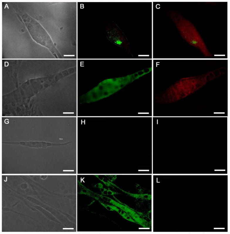
Figure 3.

Expression of the phosphorylated and unphosphorylated forms of the ErbB2 receptor in Nrg1-B-1 treated and untreated cultures. (A-C) Nrg 1-β-1 treated myotube, (A) phase contrast image of a bag fiber, (B) phospho-ErbB2 immunostaining shows an activated receptor cluster, (C) BA-G5 + phosphor-ErbB2 merge image, (D-F) Nrg 1-β-1 treated myotube, (D) phase contrast image, (E) unphosphorylated ErbB2 staining, (F) BA-G5 + unphosphorylated ErbB2 merge image, (G-I) myotube from untreated culture, (G) phase contrast image, (H) phosphor-ErbB2 staining shows no receptor activation, (I) BA-G5 staining shows no immunoreactivity, (J-L) untreated myotubes, (J) phase contrast, (K) unphosphorylated ErbB2 immunostaining shows reactivity in untreated myotube cultures, (L) BA-G5 staining is absent in non-intrafusal myotubes. Scale bars = 40μm.
