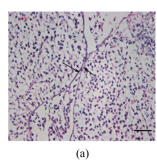
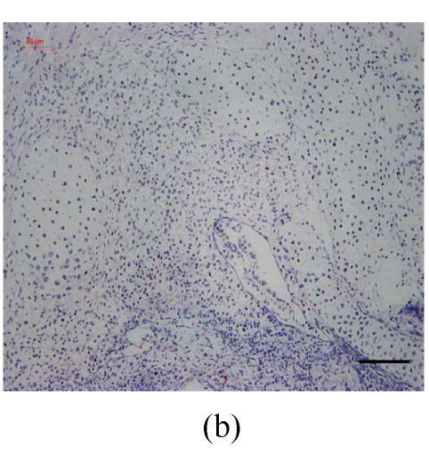
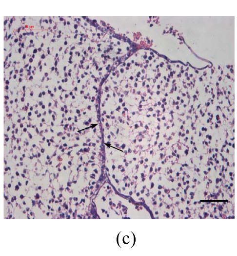
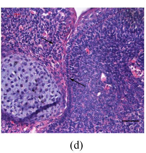
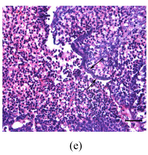
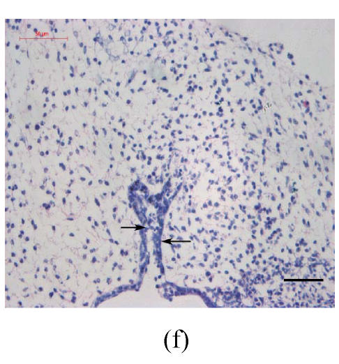
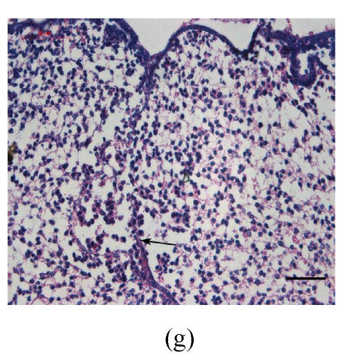
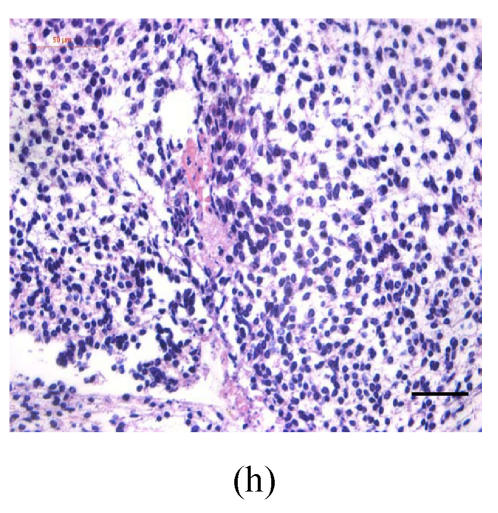
Fig. 1.
Hematoxylin and eosin (H & E)-stained sections of the palatal shelves. In the control group at 24 h of culture (a), the MEE was observed, and at 48 h (b), the MEE disappeared and the bilateral palatal shelves fused. In Dex group at 24 h (c) and 48 h (d), the MEE was still seen and after 48 h the MEE thickened. In vitamin B12-treated group at 24 h (e) and 48 h (f), the MEE also disappeared. In Dex+vitamin B12 group at 24 h (g) and 48 h (h) the bilateral palatal shelves fused
Black arrows show the MEE; Scale bars=50 µm

