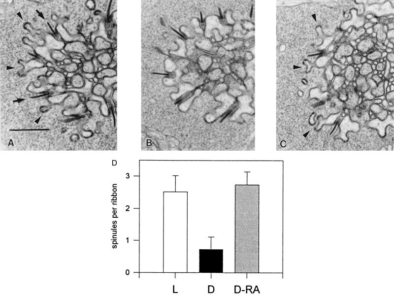
Figure 1.
Effect of at-RA on spinule formation. (A–C) Electron micrographs of tangential sections at the level of the outer plexiform layer of the carp retina. Part of a cone pedicle with ribbon synapses (arrows) is shown in each micrograph. (Bar = 1 μm.) (A) Numerous spinules (arrowheads) are visible in a light-adapted control. (B and C) Micrographs from the left and right eyes of the same dark-adapted animal. Spinules are not present in the untreated retina (B) but are present in the retina treated with at-RA (C). (D) Quantification of the spinule formation reveals that injection of at-RA into the eyes of dark-adapted animals (D-RA; n = 5 animals) results in a number of spinules significantly different from the number of spinules in the untreated eyes (D) and similar to that obtained by light adaptation (L).

