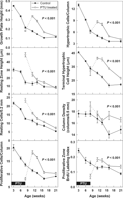
Figure 3.
Growth plate structure and function during and after PTU treatment. Female rats were made hypothyroid by adding PTU to the drinking water from birth until 8 wk of age (open symbols). Untreated animals served as controls (closed symbols). All rats received depot leuprolide acetate every 6 wk, starting at 3 wk of age. The solid boxes represent time animals received PTU (0–8 wk of age, pertains to all graphs). The dotted line represents the transition period immediately after cessation of PTU. In control animals, all end points declined significantly with age (P < 0.001). In animals that had previously received PTU, this age-dependent decline was significantly delayed for all of these end points (P < 0.001). Quantitative histology (mean ± sem) was performed on Masson Trichrome-stained sections of the proximal tibial growth plate. An observer blinded to treatment and age measured growth plate height, resting zone height, number of resting zone chondrocytes per 0.2 mm growth plate width, number of proliferative and hypertrophic chondrocytes per column, terminal hypertrophic cell height, column density, and proliferative index in the proliferative zone. Proliferative index was measured by administering BrdU to the rats 12 and 2 h before the animals were killed and then identifying BrdU-labeled cells by immunohistochemistry. The proliferative index represents the number of labeled nuclei divided by total nuclei in intact chondrocyte columns.

