
Fig. 6.
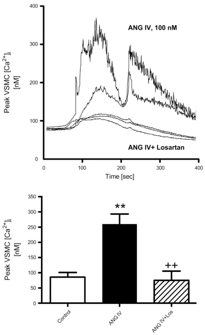
Effects of ANG IV (100 nM) on intracellular Ca2+ concentration ([Ca2+]i) in renal microvascular smooth muscle cells (VSMCs) determined using fura 2 ratiometric Ca2+ imaging (340/380). Top: time-dependent [Ca2+]i responses to ANG IV stimulation in two representative VSMCs and three other cells pretreated with losartan (10 μM) before stimulation by ANG IV. Bottom: averaged peak [Ca2+]i responses in renal VSMCs treated with perfusate only (time control), ANG IV, or ANG IV in the presence of losartan. Ratiometric [Ca2+]i imaging (340/380) was recorded continuously at 3-s intervals for up to 10 min (n = 8–10 cells/group). P < 0.01 vs. control (**) and vs. ANG IV (++).