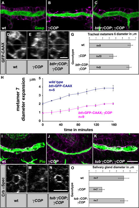
Figure 1. γCOP is required for efficient lumen diameter expansion.
(A–F, I–N) Confocal micrographs of early stage 16 wild type (A, D, I, L), γCOP mutant (B, E, J, M) and btl>γCOP; γCOP/γCOP P1 (C, F) or tub-γCOP; γCOP/γCOP P1 embryos (K, N). GFP stainings of btl>GFP-CAAX embryos reveal tracheal DT cells (A–C, in magenta and D–F, in white). Anti-Crumbs and anti-α-Spectrin stainings show SG cells (I–K, in magenta and L–N, in white). Embryos were stained for the luminal antigens Gasp (A–C) and Piopio (I–K) (green). All micrographs show confocal sections except for (D–F) and (L–N) which show yz-confocal sections of DT and SG. (G and O) Lumen diameter of DT metamere 6 using btl>GFP-CAAX (G) and diameter of SG measured for Crumbs stainings (O). γCOP mutant embryos show significantly (p<0.001) narrower trachea and SG tubes. Transgenic expression of γCOP specifically in the trachea (C, F) or under the tubulin1α promoter for SG (K, N) largely rescues the narrow lumen phenotype of γCOP mutant embryos. (H) Plot showing DT diameter expansion of live wild type and γCOP embryos expressing btl>GFP-CAAX. Embryos were recorded from late stage 13 to stage 15. Error bars are means±SD. Scale bars are 10 µm (A–C, I–K), 5 µm (D–F) and 10 µm (L–N).

