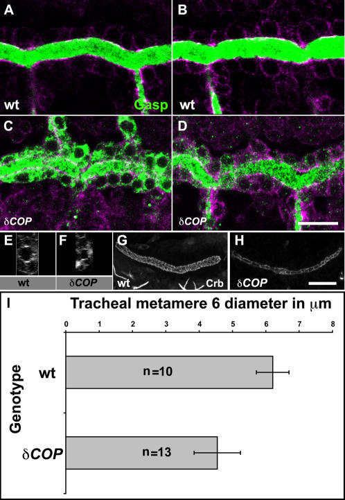
Figure 4. δCOP mutants show defects in secretion and luminal diameter expansion.
(A–H) Confocal micrographs show the DT in wild type (A, B) and δCOP mutants embryos (C, D). Embryos were stained with anti-Gasp (green) together with anti-Crumbs and anti-α-Spectrin to show tracheal cells (magenta). All micrographs are single confocal sections except (E–H) which represent yz- confocal projections of DT of metamere 6 for stage 16 wild type (E), δCOP (F) and SG projections of wild type (G), δCOP (H). δCOP mutant embryos retained a considerable amount of Gasp inside tracheal cells at stage 15 (C). At early stage 16, δCOP embryos (D, F) show narrower DT lumen in comparison to wild type (B, E). δCOP mutant embryos stained for Crb (H) show narrow SG lumen compared to wild type (G) at stage 16. (I) Graph showing the lumen diameter of the DT at metamer 6 in wild type and δCOP embryos at early stage 16 using Crb staining to visualize apical cell membrane. δCOP mutant embryos show a significant reduction in lumen diameter (p<0.001) when compared to wild type. Error bars are means±SD. Scale bars are 10 µm.

