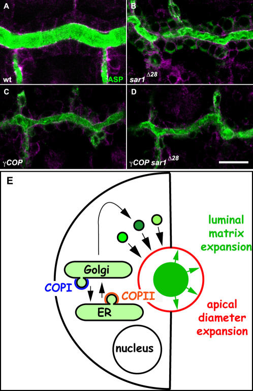
Figure 7. COPI and COPII function in tube size control.
(A–D) Confocal sections of early stage 16 wild type (A), sar1 Δ28 (B), γCOP (C) and sar1 Δ28 γCOP double mutant embryos (D). Cells of tracheal DT stained for anti-Crumbs and anti-α-Spectrin shown in magenta (A–D) and for the tracheal luminal antigen Gasp (A–D) (green). (E) Schematic illustration of a cross-section through an epithelial tube. The ER, Golgi and post-Golgi vesicles carry secreted proteins (green) into the lumen. Both COPI and COPII vesicular transport between the ER and Golgi are required for intraluminal matrix assembly and apical membrane addition (red). Green arrows indicate the proposed pressure exerted on the cells by the expanding matrix. Scale bars are 10 µm.

