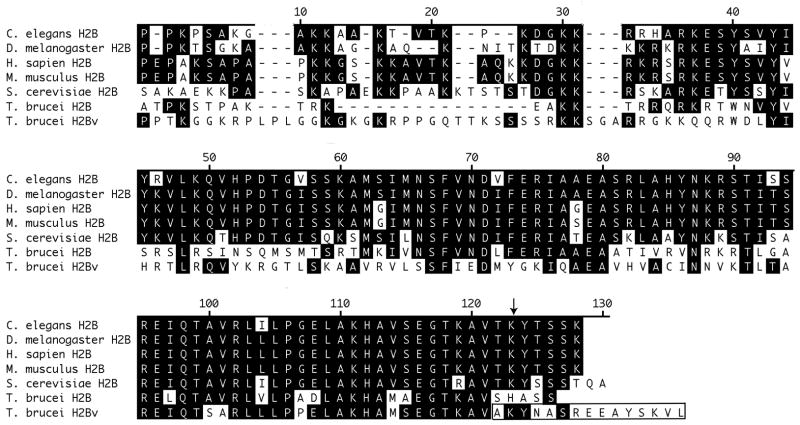
Fig. 1.
Sequence alignment of T. brucei histones H2B and H2BV with H2B from other organisms. Identical residues are shaded. The sequence ruler is numbered according to the S. cerevisiae sequence. An arrow points to T. brucei H2BV lysine 129, which is homologous to the ubiquitinated lysine 123 from S. cerevisiae. H2BV residues 128–142 are boxed.

