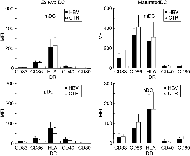
Fig. 1.

Phenotype of isolated myeloid or plasmacytoid dendritic cells (DC) analysed directly ex vivo or after 24 h of in vitro maturation with interleukin (IL)-1β, tumour necrosis factor-α, IL-6 and prostaglandin E2. Mean fluorescence intensities and standard deviations after staining for CD83, CD86, human leucocyte antigen D-related, CD40 and CD80 are shown of DC from 17 chronic hepatitis B virus carriers and controls each; none of the differences reached statistical significance.
