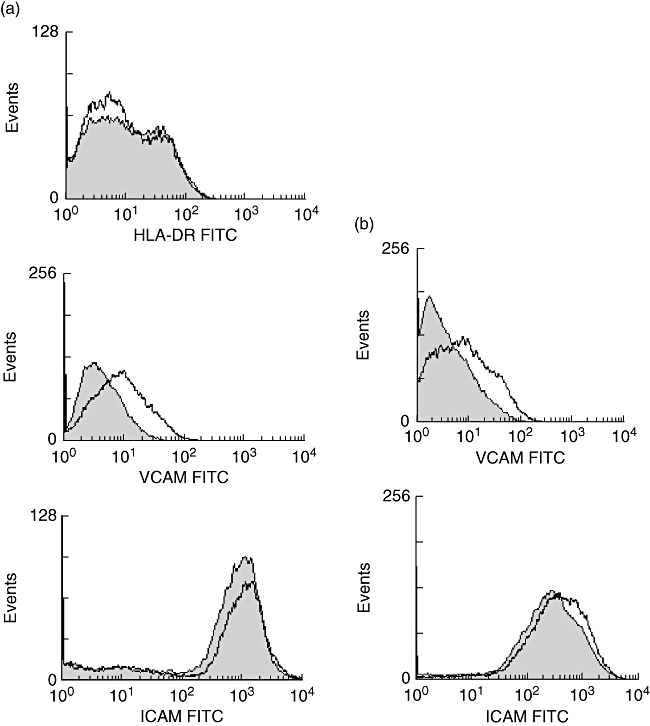
Fig. 6.

Induction of human leucocyte antigen D-related (HLA-DR) and adhesion molecules on endothelial cells. (a) Phorbol myristate acetate (PMA)/ionomycin-stimulated peripheral blood mononuclear cells were cultured for 3 days in the presence or absence of 10 μM imatinib (IM). Hereafter the supernatants were collected and added to cultured human umbilical vein endothelial cells (HUVEC) for 48 h. The expression of HLA-DR, vascular cell adhesion molecule (VCAM) and intercellular adhesion molecule (ICAM) was assessed by fluorescence activated cell sorter (FACS) analysis in HUVEC incubated with supernatants of IM/PMA/ionomycin-treated T cells (filled histogram) and in HUVEC incubated with supernatants of PMA-ionomycin-treated T cells (open histogram). (b) HUVEC were stimulated for 3 days with 10 ng/ml of tumour necrosis factor-α. The cells were cultured in the presence or absence of 10 μM IM. Hereafter, the surface expression of ICAM and VCAM was determined by FACS analysis. The filled histogram represents antigen expression in the absence of IM, the open histogram represents antigen expression in the presence of IM. In (a) and (b) the results of a representative experiment (n = 3) are depicted.
