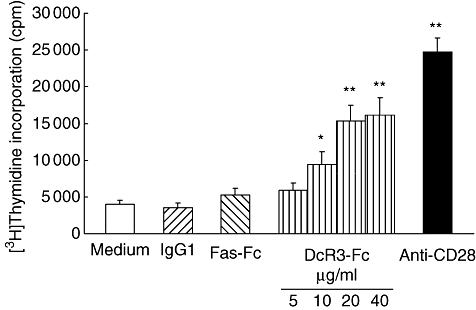
Fig. 3.

Soluble decoy receptor 3 (DcR3)–Fc enhances human T cell proliferation in conjunction with suboptimal concentration of immobilized anti-CD3. Purified human T cells from peripheral blood mononuclear cells were cultured for 72 h in 96-well flat-bottomed microtitre plates precoated with an anti-human CD3 monoclonal antibody (mAb) (500 ng/ml) and soluble DcR3–Fc recombinant protein (5–40 μg/ml), soluble Fas–Fc recombinant protein (10 μg/ml) anti-CD28 mAb (1 μg/ml) or human immunoglobulin G1 (IgG1) (10 μg/ml) as controls. The cultures were pulsed with [3H]-thymidine (1·0 μCi/well) 18 h before harvesting the cells, and [3H]-thymidine incorporation was measured. The mean ± standard deviation of the counts per minute value from triplicate samples is shown. The experiments were performed more than three times, and a representative data set is presented. Statistical analysis by Student's t-test revealed significant differences between human immunoglobulin 1- and soluble DcR3–Fc-treated samples (*P < 0·05; **P < 0·01). A similar result was obtained with T cells purified from 20 other independent healthy donors.
