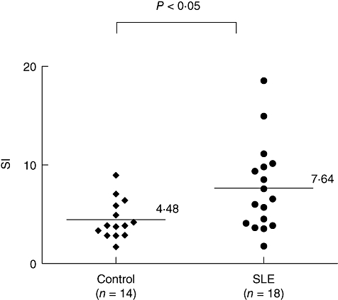
Fig. 5.

Enhanced T cell reactivity to decoy receptor 3 (DcR3)-induced co-stimulation in T cells from patients with systemic lupus erythematosus (SLE). Purified T cells from peripheral blood mononuclear cells of SLE patients were cultured for 72 h in 96-well flat-bottomed microtitre plates precoated with an anti-human CD3 monoclonal antibody (500 ng/ml) and soluble DcR3–Fc recombinant protein (10 μg/ml) or human immunoglobulin G1 (IgG1) (10 μg/ml) as controls. The cultures were pulsed with [3H]-thymidine (1·0 μCi/well) 18 h before harvesting the cells, and [3H]-thymidine incorporation was measured. The stimulation index (SI) was calculated as the ratio of counts per minute (cpm) of DcR3–Fc to that of IgG1. The SI was calculated according to the formula: cpm of T cells stimulated with anti-CD3 and DcR3–Fc/cpm of T cells stimulated with anti-CD3 and IgG1. Background cpm obtained with T cells in medium alone was negligible. Each symbol represents one donor (n = 18 for SLE and n = 14 for normal healthy subjects). The mean SI obtained with these donors is indicated (P < 0·05 by the Mann–Whitney rank sum test).
