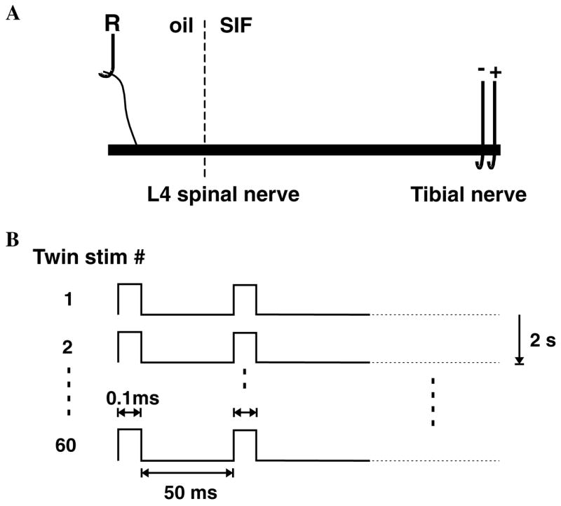
Fig. 1.

Experimental paradigm. (A) In vitro preparation. The L4 spinal nerve in continuity with the tibial nerve was excised and placed into a two chamber in vitro bath. In one oil-filled chamber, a small filament of the L4 spinal nerve was placed on a silver electrode for extracellular recording of action potential activity in single C fibers. In the other SIF perfused chamber, a suction electrode was applied to the distal end of the tibial nerve. (B) Stimulation protocol. Electrical stimulation consisted of twin pulses with an interstimulus interval of 50 ms that were applied every 2 s for a total of 60 twin pulses in the train. The latency of the electrically evoked action potential was used to measure how the conduction velocity varied with repeated stimulation.
