
Fig. 1.
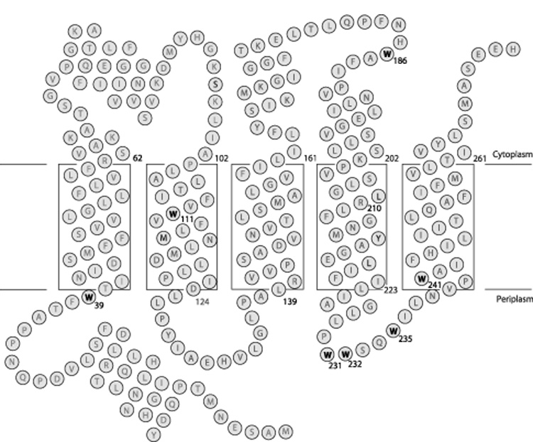
Topological model for folding of subunit a in E. coli inner membrane. The biochemical evidence for the insertion of the five TMHs is discussed in the text. The depth of placement of the helices in the membrane is based upon cross-linking studies as described elsewhere [29]. The positions of the seven Trp residues in the wild type protein are highlighted. The helical segments shown in regions peripheral to the lipid bilayer were predicted by backbone chemical shift analysis for the protein dissolved in chloroform-methanol-water solvent [32].