
Fig. 5.
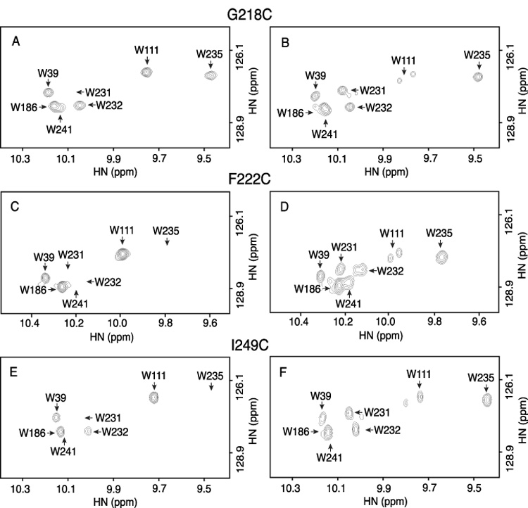
Line broadening of Trp ε-imino indole signals by PROXYL spin label attached at positions 218 (A,B), 222 (C,D) and 249 (E,F) of subunit a. Spectra of the spin labeled proteins are shown on the left (A,C,E) and the control spectra of the samples reduced with phenylhydrzine on the right (B,D,F). Resonance assignments are indicated by the arrows.