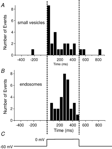
Figure 4.

Times of fusion relative to a 0.5 s depolarization A, timing of exocytosis of presumptive synaptic vesicles loaded using protocol 1 followed by a 30–60 min wash, plotted as histogram. B, endosomes labelled using protocol 2. Vertical dashed lines indicate the beginning and end of the depolarization (C). Note that endosomes, but not synaptic vesicles, fuse after a delay.
