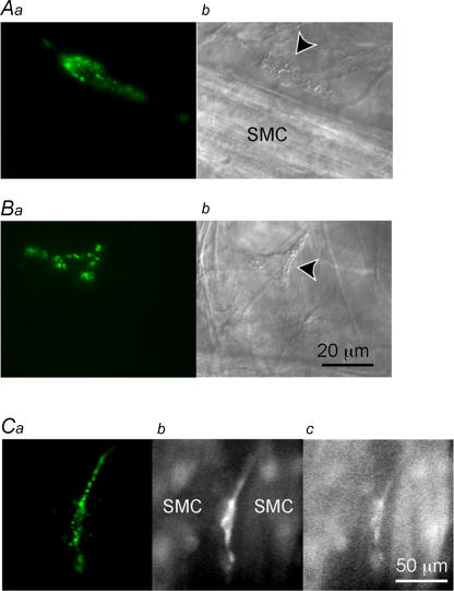
Figure 1. Identification of ICC-LCs in the rabbit urethra Panels.
a show fluorescent images of ICC-LCs in the rabbit urethra stained using ACK2 antibody against Kit labelled with Alexa 488. Panels b show micrographs of preparations viewed with Nomarski optics. A, ICC-LC (arrow head) which had a spindle-shaped cell body is shown lying in parallel with a muscle bundle (SMC). B, another ICC-LC having a stellate-shaped cell body is shown located in the connective tissue between the muscle bundles. C, in a different preparation, which had been loaded with fura-2, ICC-LCs identified by immunoreactivity against Kit (a) had higher F340 fluorescence than neighbouring smooth muscle cells (SMC, b), whilst having similar F380 fluorescence (c).

