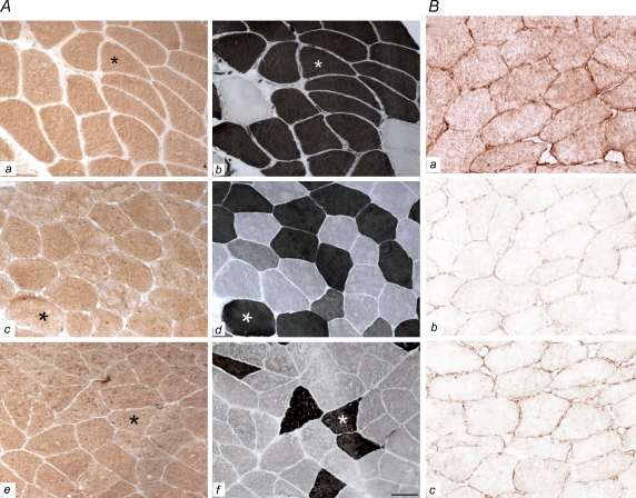
Figure 2. IL-15 expression.
A, IL-15 expression is shown for triceps (a), vastus lateralis (c) and soleus (e) muscles. Myofibrillar ATPase staining is shown in neighbouring tissue sections of the triceps (b), vastus lateralis (d) and soleus (f) muscles (b, d and f are consecutive sections to those shown in a, c and e). Fibre types are distinguished by ATPase staining and fibres appearing black are type 2 fibres. The asterisks depict matching fibres for each muscle group (n= 7). As displayed, IL-15 protein expression is comparable in muscle fibre types and in the different muscles of triceps, vastus lateralis and soleus. Scale bar: 89 μm. B, IL-15 expression as defined by our standard IL-15 IHC is shown in a, while negative control sections are shown in b and c. Bb, negative control section as seen after the primary antibody was pre-absorbed with its corresponding antigenic protein, thereby preventing the following IHC binding of primary IL-15 antibodies to endogenous IL-15 proteins of the muscle. Bc, negative control section after incubation in the absence of primary IL-15 antibodies, whereby the IHC results in negative immunostaining. Scale bar: 47 μm

