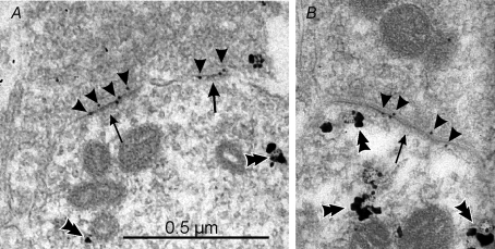
Figure 4. Renshaw cells express postsynaptic receptors that include GluR2 and GluR4 subunits in their composition.
The images show calbindin-IR dendrites sampled from the ventral Renshaw cell region, immunostained in pre-embedding with immuno-nanogold and amplified with silver (double arrowheads). A proportion of postsynaptic densities (arrows) at excitatory synapses can be labelled with post-embedding immunogold (10 nm, arrowheads) for GluR4 AMPA receptor subunits in A and B. Similar results were obtained with antibodies against GluR2/3 or specific for GluR2. Although the combined pre- and postembedding immunocytochemistry and cryosubstitution techniques necessary to reveal these receptor subunits results in slightly compromised ultrastructure, particularly vesicle size and shape, the PSDs are asymmetric and contain AMPA receptors; hence, they are highly likely to represent excitatory (Type 1) synapses. Antibodies against GluR1 did not immunolabel any synapses on Renshaw cells, but intensely immunolabelled synapses in the dorsal horn. The images were obtained from a P20 animal.

