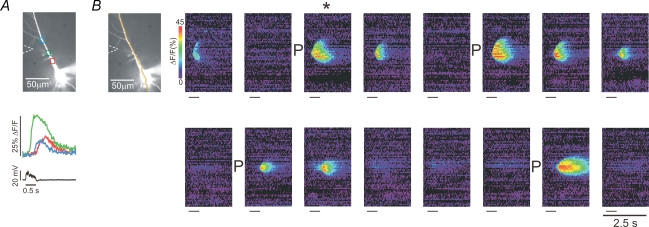
Figure 1. Priming stores with repetitive action potentials prepares pyramidal neurons to release calcium.
A, typical response of a neuron to tetanic stimulation. The image shows a cell filled with bis-fura-2 with three ROIs indicated. The cell was filled with indicator from the patch pipette on the soma, which also recorded the electrical response. The position of the stimulating electrode is also shown. Following tetanic stimulation (50 pulses at 10 ms intervals; 90 μA current for 100 μs), delayed [Ca2+]i increases were detected at the three locations. This event is the trial indicated with * in the second part of the figure. B, response to the same stimulation protocol at 2 min intervals. The pseudocolour images show a ‘line scan’ along the pixels indicated in the cell image. When Ca2+ release was evoked it occurred earliest at a location in the dendrites and spread as a wave in a restricted region of the cell. At four times during the experiment (indicated by ‘P’ next to the images) action potentials were evoked intrasomatically at 4 Hz for the duration of the 2 min intertrial interval. Following this priming protocol Ca2+ release was strongest. The short lines under each image indicate the time of tetanic stimulation. The 2.5 s scale bar applies to all the images.

