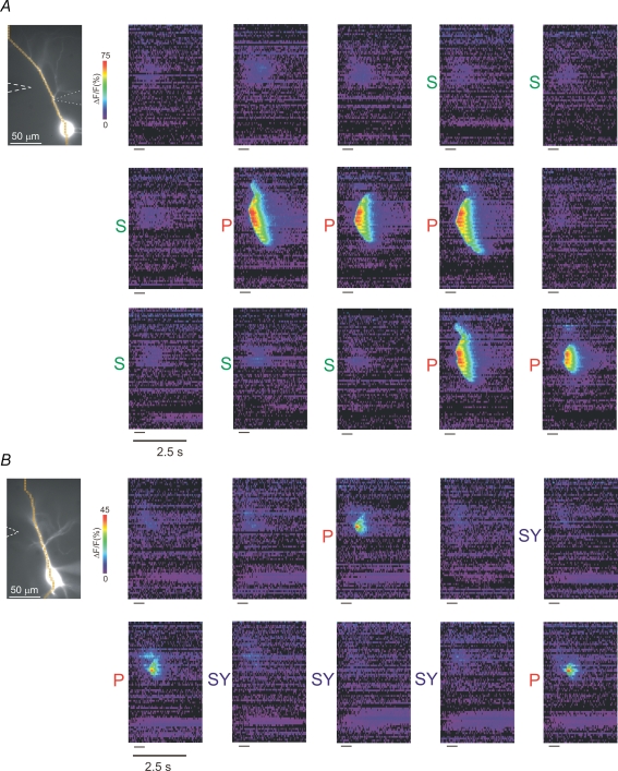
Figure 4. Priming with action potentials is more effective than priming with a sustained depolarizing pulse (SSD) or repetitive subthreshold synaptic stimulation (SY).
A, SSD. The cell on the left was patched on the main apical dendrite about 50 μm from the soma. The pseudocolour images are ‘line scans’ of successive trials with an intertrial interval of 15 s, ordered sequentially. The horizontal line on the lower left of each image represents the tetanic synaptic stimuli (50 pulses at 10 ms intervals, 40 μA). The white dashed arrow on the image indicates the position of the stimulating electrode. The scale bar of 2.5 s applies to all the images. The images preceded by a P represent trials in which the cell was primed with action potentials at 4 Hz during the intertrial interval before the cell was stimulated synaptically. The images preceded by an S represent trials in which the cell was injected with a constant current pulse to bring the membrane potential just below the threshold for spike firing (2 mV below threshold in this example) during the intertrial interval. Successful Ca2+ release occurred in trials primed with action potentials while there were no trials with successful release in trials primed with sustained subthreshold depolarization. B, SY. The images preceded by SY represent trials in which the cell was stimulated synaptically during the 15 s intertrial interval at 0.5 Hz with the same tetanic stimulus train used for evoking release (30 μA) except at a lower current (20 μA), which was below the threshold for spike firing and did not elicit a Ca2+ release wave. Successful Ca2+ release occurred in trials primed with action potentials at 0.5 Hz while there were no trials with successful release in trials primed with repetitive synaptic stimulation.

