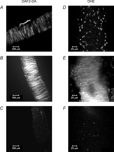
Figure 3. Imaging of nitric oxide and superoxide anion availability in intact resistance arteries.
Extended focus reconstructions obtained with Metamorph image analysis software of serial optical sections taken from a rat mesenteric resistance artery stained with DAF2-DA (left panels, ×20 air objective) or dihydroethidium (DHE) (right panels, ×63 oil immersion objective) and fixed after staining. A, DAF2 emitted fluorescence in non-stimulated conditions (basal NO). B, DAF2 emitted fluorescence after stimulation with 10−6m acetylcholine (stimulated NO). C, DAF2 emitted fluorescence after pre-incubation with the NO synthase inhibitor l-NAME (negative control). D, ethidium bromide emitted fluorescence in non-stimulated conditions (basal superoxide anion). E, ethidium bromide emitted fluorescence in an artery stimulated with 10−6m pyrogallol (stimulated superoxide anion). F, ethidium bromide emitted fluorescence in an artery pre-incubated with superoxide dismutase (negative control).

