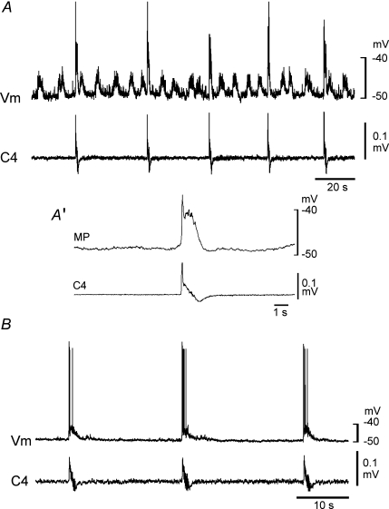
Figure 3. Inspiratory neurons in the ventrolateral medulla of Atp1a2−/− mice.
Burst activity was induced by application of 5 μm adrenaline. A, an inspiratory neuron receiving clustered excitatory postsynaptic potentials (EPSPs). A′, membrane potential averaged 24 times with the use of C4 activity as a trigger. B, an inspiratory neuron receiving neither clustered EPSPs nor clustered inhibitory postsynaptic potentials (IPSPs). Note the absence of clear EPSPs associated with inspiratory bursts. The threshold for action potential generation was approximately −40 and −35 mV in the neuron in A and the neuron in B, respectively. Vm, membrane potential trajectory; C4, C4 inspiratory activity.

