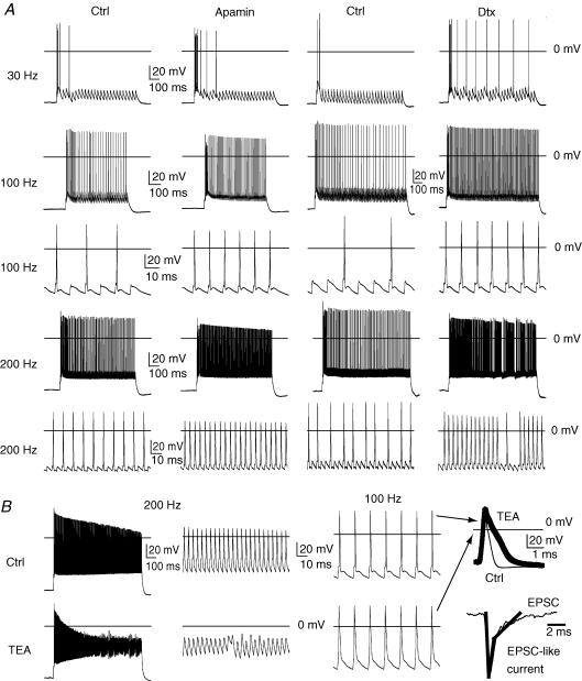
Figure 8. Role of Kv1, SK and Kv3.2 in the firing response to excitatory postsynaptic currents (EPSCs).
EPSC stimuli were designed based on the kinetics of EPSC events observed in these neurons during whole-cell voltage clamp (−70 mV, Fig. 8B bottom right). The current pulse used to model an EPSC stimulus is shown in the thicker tracing overlay with peak current reached in 0.5 ms and decaying to 40% of peak value in 1 ms. A slower component decays to baseline over a 5 ms period. A, EPSC-like stimuli at 30 Hz (1000 pA peak current) caused an initial burst of action potentials and then largely failed to evoke firing in controls (Ctrl). Washing in apamin (300 nm) to inhibit SK Ca2+-activated K+ channels in this same control neuron increased the number of spikes within the burst and improved coupling over the first few EPSC-like stimuli. Washing in α-dendrotoxin (100 nm) to block Kv1 K+ channels increased the number of spikes within the burst, but also increased EPSC-spike coupling. Bath application of apamin and α-dendrotoxin increased EPSC-spike coupling at 100 Hz (900 pA peak) and 200 Hz (700 pA peak). B, washing in the non-selective Kv3.2 channel blocker (TEA, 1 mm) depolarized the interstimulus membrane potential and attenuated action potential height with 200 Hz EPSC-like stimuli (900 pA, peak). At 100 Hz (900 pA, peak), action potentials at 100 Hz were widened, but only weakly attenuated, by 1 mm TEA (far right, thick trace versus Ctrl, thin trace, 1 ms and 20 mV scale).

