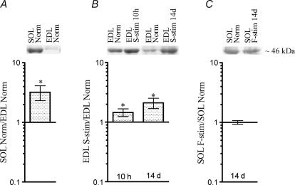
Figure 5.
Levels of T115-phosphorylated MyoD differ between EDL and soleus, and are influenced by activity The level of MyoD T115-phosphorylated protein from nuclear extracts quantified on Western blots stained with the anti-pMyoDT115 antibody in normal solei relative to EDL (A), in normal EDL stimulated with a slow pattern (S-stim) 10 h and 14 days relative to normal EDL (B), and in soleus muscles stimulated with a fast pattern (F-stim) for 14 days relative to normal solei (C). Data are given as mean ±s.e.m. on a logarithmic scale, n= 6–11, and as representative Western blots (upper panels). *P≤ 0.05. Numerical values are given in Table S1.

