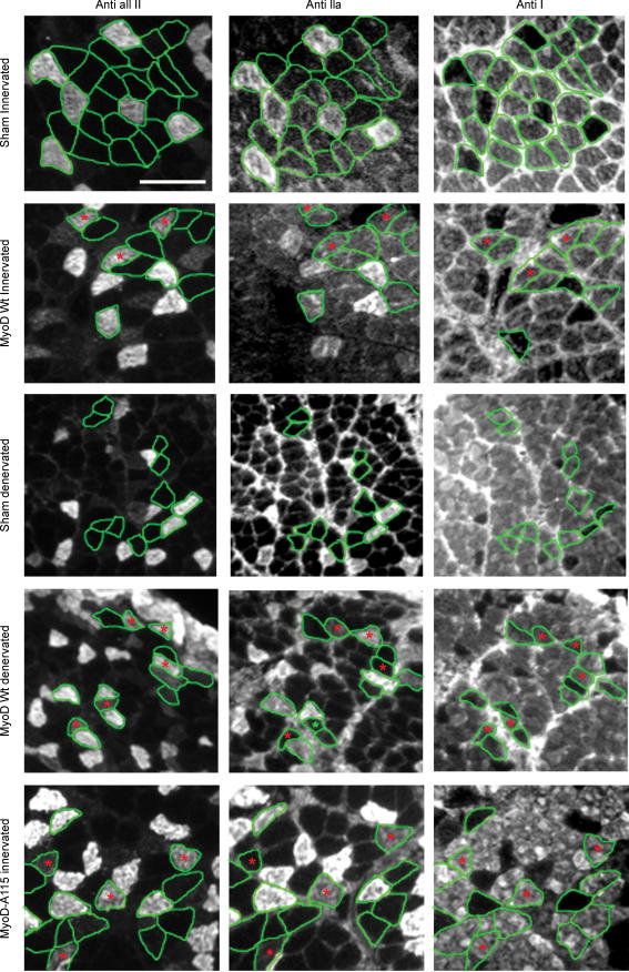
Figure 6.
MyHC immuno-histochemistry on neighbouring cross sections from rat soleus Transfected fibres are encircled in green and hybrid fibres are marked with red asterisks. Notice the increased amount of hybrid fibres in MyoD-positive denervated fibres and T115A-mutated MyoD fibres. All fibres were negative for IIb MyHC (not shown). Scalebar, 100 μm.

