
Figure 5.
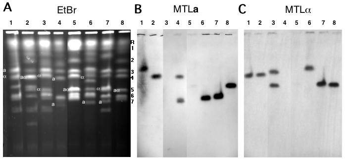
Candida dubliniensis pulse-field chromosome separations. A. CHEF separation of chromosomes from C. albicans strain 1006 and from 7 C. dubliniensis strains. Three lanes from the middle of the gel which contained irrelevant strains have been removed from the figure. The letters to the right of this figure indicate the positions of the C. albicans chromosomes used as molecular markers: 1, 3.2 Mb; 2, 2.2 Mb; 3, 1.8 Mb; 4, 1.6 Mb; 5, 1.2 Mb; 6, 1.03Mb; 7, 1.0 Mb. Chromosome R, as mentioned above, does not migrate strictly according to its molecular size in a CHEF gel. B. Southern blot probed with a sequence from the C. dubliniensis MTLa1 gene. C. Southern blot probed with a sequence from the C. dubliniensis MTLα1 gene. Lane 1, Strain R3b; 2, R1b; 3, 3225; 4, 3233; 5, 1006 (C. albicans); 6, CD36; 7, 16F; 8, 514.