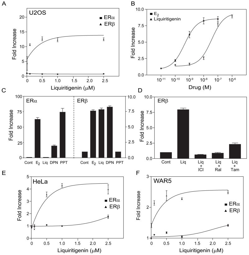
Fig. 2.
Liquiritigenin selectively activates transcription through ERβ. (A) ERE tk-Luc was cotransfected into U2OS cells with expression vectors for ERα or ERβ. After transfection, the cells were treated for 18 h with liquiritigenin and luciferase activity was measured. Dose-response curves for E2 and liquiritigenin in U2OS cells. Following transfection the cells were treated with increasing amounts of E2 or liquiritigenin for 18 h (B). ERE tk-Luc was cotransfected into U2OS cells with expression vectors for ERα (left panel) or ERβ (right panel) and then treated with 10 nM E2, 1 μM liquiritigenin, 1 μM DPN or 1 μM PPT for 18 h and then luciferase was measured (C). The activation by liquiritigenin is blocked by anti-estrogens. (D) ERE tk-Luc was cotransfected into U2OS cells with an expression vector ERβ and the cells were treated with 1 μM liquiritigenin in the absence or presence of 1 μM ICI 182780 (ICI), raloxifene (Ral) or tamoxifen (Tam). Liquiritigenin selectively activated the ERE tk-Luc with ERβ in HeLa cervical (E) and WAR5 prostate cancer (F) cell lines. Each data point is the average of triplicate determinations ± S.E.M. An activation by the drug was significant (p < 0.05) when it was 2-fold greater than the control values.

