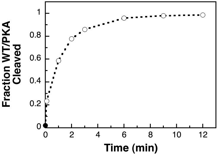
Figure 3.
Rapid cleavage of WT/PKA with excess RecA. A representative time-course shows the cleavage of 0.5 μM WT/PKA in the presence of 1 μM activated RecA (open circles) at 30°C. The first time-point was stopped 6 s after adding RecA, and was 23% cleaved. Prior to adding RecA, the WT/PKA was 2% cleaved (solid circle).

