
Figure 1.
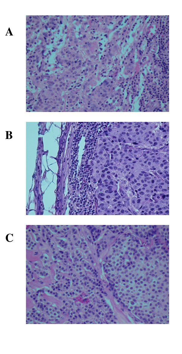
Hematoxylin and eosin stained slides of formalin-fixed, paraffin-embedded tissue sample blocks of breast tumor tissue and metastatic breast tumor tissue in lymph node. Panel A (father): metastatic ductal carcinoma of breast in axillary lymph node. The tumor is almost completely replacing the normal tissue in this 2-cm node. Note the rim of residual subcapsular lymphoid tissue. Panel B (mother): invasive moderately differentiated ductal carcinoma of breast. Note the prominent lymphocytic response. Panel C (daughter): invasive and in situ lobular carcinoma of breast. Only a portion of the round edge of a lobule containing lobular carcinoma in situ is seen here. Magnification is 200X.