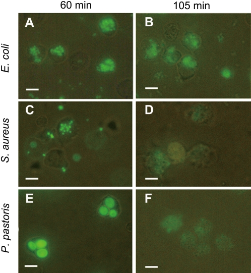
Figure 4. Phagocytosis of E. coli, S. aureus and P. pastoris by macrophages.
Vg, FITC-labeled microbes and macrophages freshly isolated were mixed and incubated together at room temperature. The controls were performed in the presence of BSA instead of Vg and in the absence of Vg. Phagocytosis was observed at 60 min after mixing macrophages with microbes. (A, C and E) Phagocytosis of E. coli, S. aureus and P. pastoris by macrophages at 60 min; (B, D and F) Phagocytosis of E. coli, S. aureus and P. pastoris by macrophages at 105 min. Scale bars: 10 µm.

