
Fig. 3.
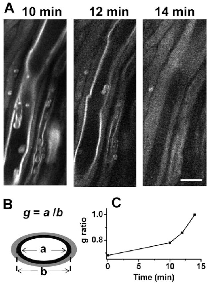
Real-time CARS imaging of myelin degradation. A: Time-lapse CARS images of myelin swelling in the spinal tissue incubated with a Krebs’ solution containing 10 mg/ml lyso-PtdCho. B: Diagram of measuring the g ratio of a partially swollen myelin fiber based on the remaining compact region. C: The increase of g ratio during the process of myelin swelling. Scale bar = 10 μm.