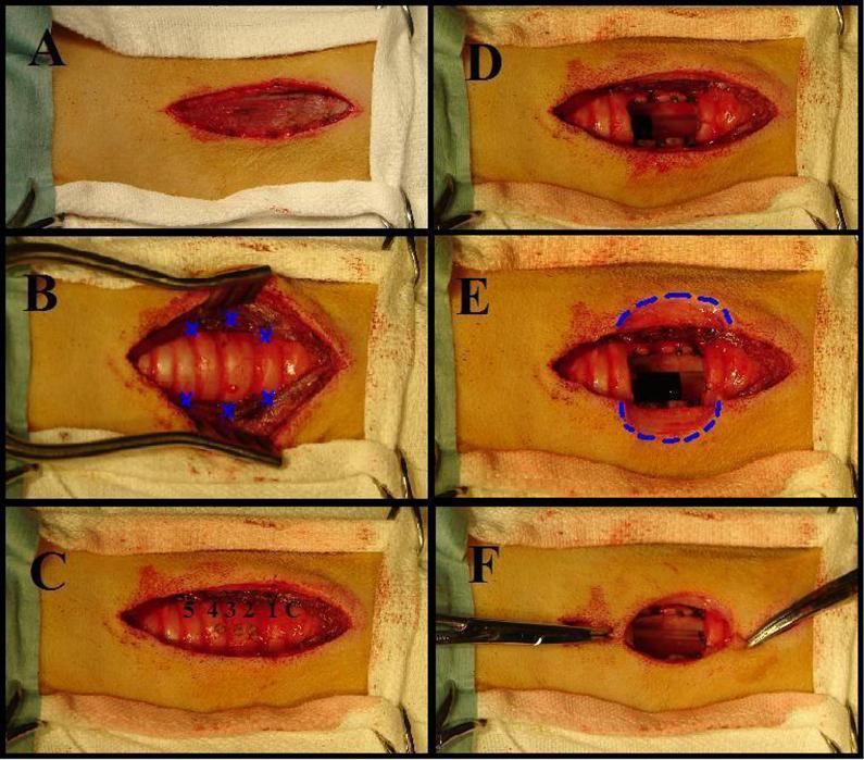
Figure 1.
A: A 6 cm incision is made parallel to the trachea on the anterior neck. B: The trachea is exposed and the sternohyoid muscles are sutured to the lateral tracheal wall. Blue X’s mark the location of sutures. C: Tracheal rings 2,3,4 are identified and marked with electro-cautery. D: Rings 2,3,4 are removed creating a rectangular stoma, 2 × 1 cm in the anterior wall of the trachea. E: A four-leafed clover shaped incision was formed by removing a 2 cm × 1 cm semicircle of skin from either side of the tracheal opening (Dotted Blue Line). F: The edges of the four-leafed clover shaped incision approximate to the edges of the stomal opening.

